Clinical, Radiological features of Calcifying Epithelial Odontogenic Tumor: Report of two rare cases
Calcifying Epithelial Odontogenic Tumor
DOI:
https://doi.org/10.56501/intjorofacres.v6i2.507Keywords:
CEOT, Computed Tomography, Calcifications, Diagnosis, Pindborg tumorAbstract
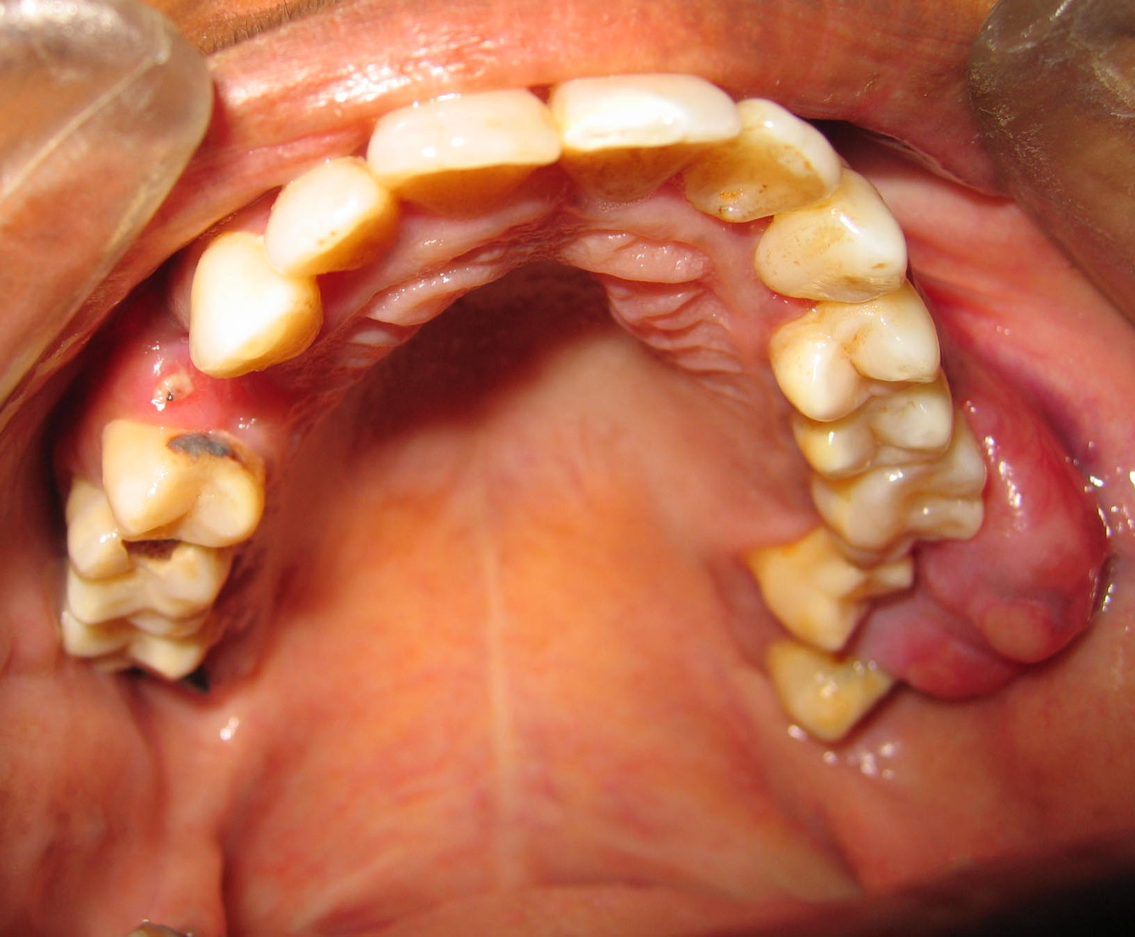
The calcifying epithelial odontogenic tumor (CEOT) is a rare benign odontogenic tumor constitutes around 1% of all odontogenic tumours involving the jaw. The intraosseous and extraosseous variant constitutes about 95% and 5% of CEOT respectively. We have reported two CEOT cases. One is associated with an impacted left third molar in the maxilla of 39-year-old female patient, and the other with a sessile soft tissue gingival mass in the left molar region of a 30-year-old female patient. It also emphasizes the importance of advanced imaging and peculiar findings of computed tomography in diagnosing this rare tumour. The visualization of the internal structure of the lesion and the involvement of the neighbouring structures were considered very helpful for diagnosis and treatment planning. Early detection and treatment planning for such rare cases is required to prevent further complications.
Publication Facts
Reviewer profiles N/A
Author statements
Indexed in
- Editor & editorial board
- profiles
- Academic society
- N/A
- Publisher
- MM Publications

To learn about these publication facts, click ![]()
PF is maintained by the Public Knowledge Project
Downloads
Published
How to Cite
Issue
Section
License
Copyright (c) 2022 Dr. Om Dnyanadev Kharat

This work is licensed under a Creative Commons Attribution-NonCommercial 4.0 International License.


